|
Jason J. Corso
|
Snippet Topic: Medical Imaging
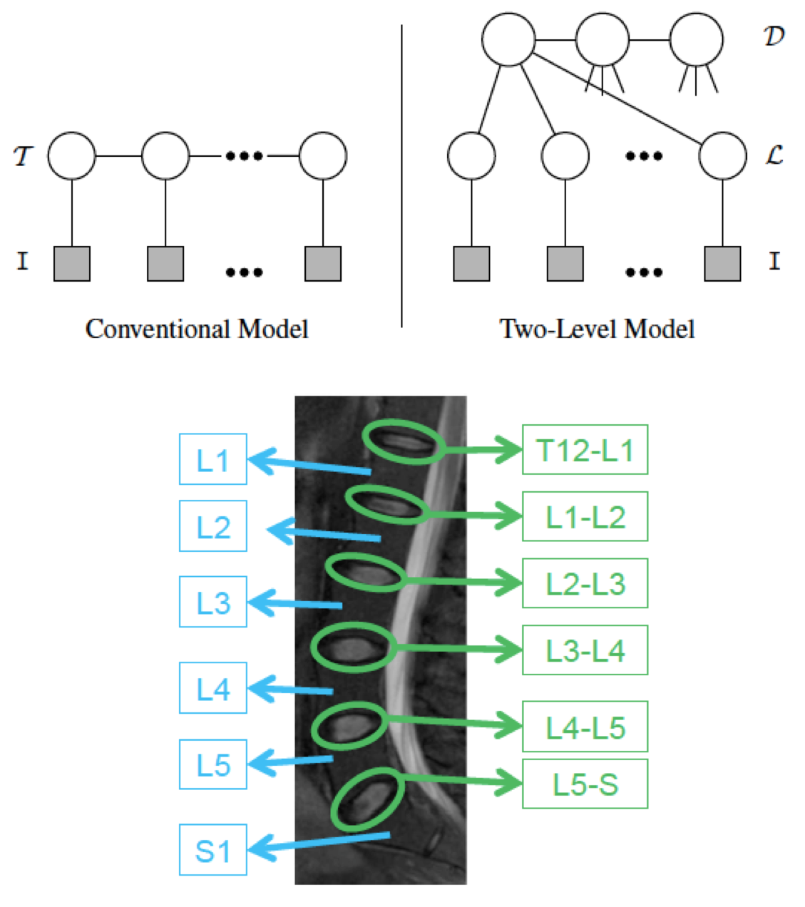
Two-Layer Probabilistic Model Jointly on Pixel and Object Features  | We have developed a two-level probabilistic model for jointly localizing and labeling lumbar discs Whereas conventional labeling approaches (e.g., our approach to brain tumor above) define all models at the pixel level, our model integrates both pixel-level information, such as appearance, and object-level information, such as relative location and shape. Utilizing both levels of information adds robustness to the ambiguous disc intensity signature and high structure variation. Yet, we are able to do efficient (and convergent) localization and labeling with generalized expectation-maximization. We have presented accurate results, about 89% accuracy on 105 normal and abnormal cases (96% when using normal alone and 87% when using abnormal alone). More information.
|
|
R. S. Alomari, J. J. Corso, and V. Chaudhary.
Labeling of lumbar discs using both pixel- and object-level features
with a two-level probabilistic model.
IEEE Transactions on Medical Imaging, 30(1):1-10, 2011.
[ bib |
.pdf ]
|
|
|
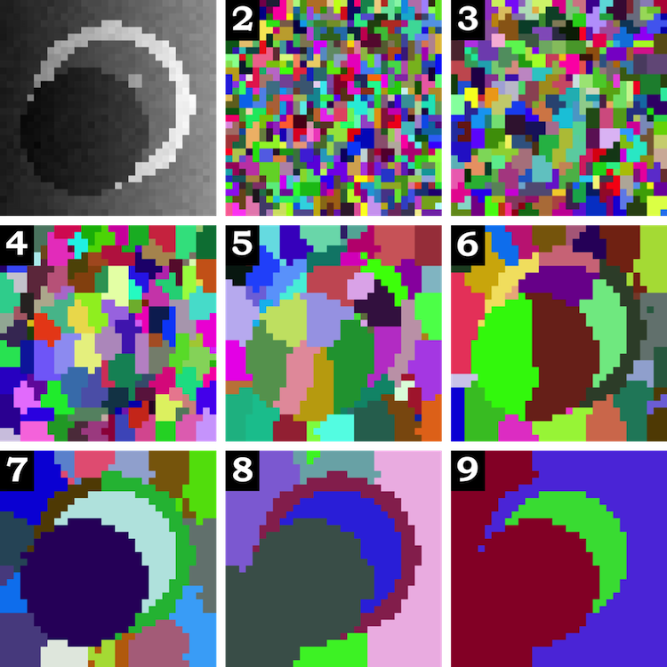
Multilevel Segmentation with Bayesian Affinities  | Automatic segmentation is a difficult problem: it is under-constrained, precise physical models are generally not yet known, and the data presents high intra-class variance. In this research, we study methods for automatic segmentation of image data that strive to leverage the efficiency of bottom-up algorithms with the power of top-down models. The work takes one step toward unifying two state-of-the-art image segmentation approaches: graph affinity-based and generative model-based segmentation. Specifically, the main contribution of the work is a mathematical formulation for incorporating soft model assignments into the calculation of affinities, which are traditionally model free. This Bayesian model-aware affinity measurement has been integrated into the multilevel Segmentation by Weighted Aggregation algorithm. As a byproduct of the integrated Bayesian model classification, each node in the graph hierarchy is assigned a most likely model class according to a set of learned model classes. The technique has been applied to the task of detecting and segmenting brain tumor and edema, subcortical brain structures and multiple sclerosis lesions in multichannel magnetic resonance image volumes. More information. |
|
|


